Human brain in 3D
Explore a network for movement, memory and sensation. Explore the 3D model, inspect its parts and learn with guided explanations. Free on Atlas.
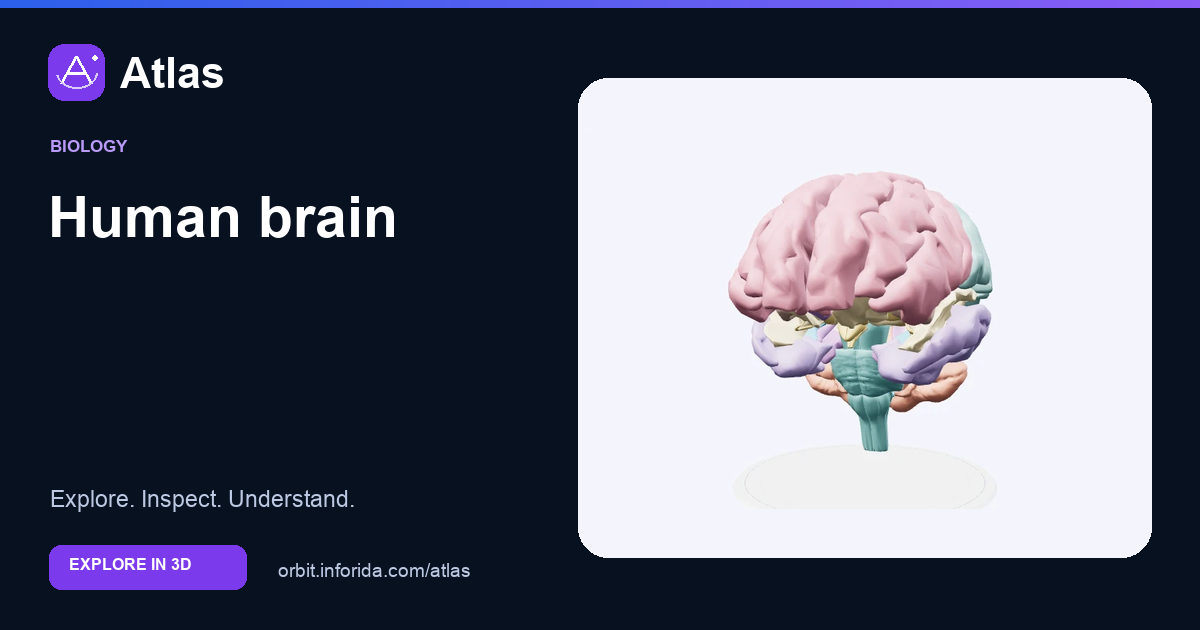
What you can learn
Connect major brain regions without assigning a whole ability to one small spot.
The cerebral hemispheres contain folded cortex and underlying white matter. Folds increase cortical surface area.
The cerebellum contributes to coordination; the brainstem links the brain with the spinal cord and supports essential functions.
Fluid-filled ventricles are spaces within the brain. They are not empty tissue or separate thinking centres.
Try an investigation
Locate the cerebellum and brainstem from two camera angles. Explain how a sensory signal and a motor command could travel through different connected regions.
Check your understanding
Which structure is especially important for coordinating movement and balance?
Cerebellum. The cerebellum helps coordinate movement and maintain balance.
Which structure links much of the brain with the spinal cord?
Brainstem. The brainstem contains pathways connecting the brain and spinal cord and supports vital functions.
What are brain ventricles?
Interconnected fluid-filled cavities. Brain ventricles contain cerebrospinal fluid. They are distinct from heart ventricles.
What is a major role of the corpus callosum?
Connect the cerebral hemispheres. Its nerve fibres support communication between regions in the two hemispheres.
Why is it misleading to assign all memory to a single spot in this model?
Memory depends on interacting brain networks. Different structures contribute to memory and learning through their connections. Model colours identify structures rather than individual memories.
Sources and model scope
Surface anatomy does not reveal thoughts, intelligence, feelings or a learner’s attention. Many functions rely on distributed networks.