Human eye in 3D
Investigate how light becomes a signal. Explore the 3D model, inspect its parts and learn with guided explanations. Free on Atlas.
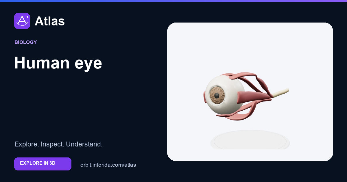
What you can learn
Separate the optical path from the neural pathway.
The cornea and lens bend light toward the retina.
The iris regulates the pupil opening, changing how much light enters; lens shape contributes to focusing.
Photoreceptors begin neural signalling in the retina. Signals travel through the optic nerve toward the brain.
Try an investigation
Compare the cornea and lens, then locate the retinal surface. Explain where optical refraction differs from neural signalling. Compare these internal tissues with the external muscles that move the eye.
Check your understanding
Which tissue contains the photoreceptors that detect light?
Retina. Retinal photoreceptors convert light into signals that are processed and sent toward the brain.
Which opening allows light into the eye?
Pupil. The pupil is the opening in the iris. Its size helps regulate incoming light.
What does the lens help do?
Focus light on the retina. The lens changes shape to help focus objects at different distances.
Sources and model scope
The default HRA reference contains 38 source ocular surfaces, separate from the legacy BodyParts3D view. An optional enlarged study compares rods and cones, simplified retinal connections, light/output orientation and the optic-disc exit. These are original cell symbols and tissue comparisons, not measured microscopic layers or a complete retinal circuit. Dynamic accommodation, optical image formation and neural physiology are not simulated; this model cannot test eyesight.